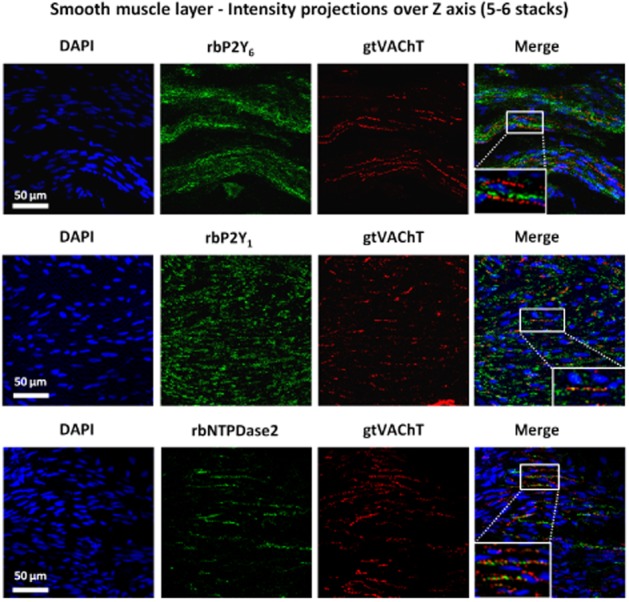
Figure 9.

Confocal micrographs showing P2Y6, P2Y1 and NTPDase2 immunoreactivity in transverse sections of the detrusor smooth layer of rat urinary bladder. To facilitate visualization of small cholinergic nerve terminals staining for VAChT (red) images correspond to the intensity projections over Z axis of five to six confocal microscopy stacks taken at the smooth muscle layer. No co-localization was found between P2Y6 receptor (green) and VAChT (red) immunoreactivity. Conversely, VAChT-positive cholinergic nerve terminals (red) stained positively with antibodies against the P2Y1 receptor and E-NTPDase2 (green); yellow staining denotes co-localization. Nucleic DNA is stained with DAPI (blue). Scale bars = 50 μm.
