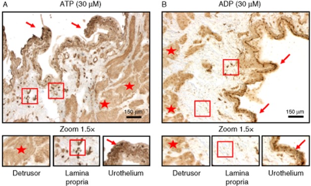
Figure 10.

Histochemical E-NTPDase activity in the rat urinary bladder. Phosphate deposition resulting from extracellular catabolism of ATP (30 μM, A) was found predominantly in the urothelium (arrows), but was also present in the sub-urothelial (square) and smooth muscle (star) layers: Extracellular ADP (30 μM, B) was dephosphorylated predominantly in apical urothelial cells (arrows) and in the smooth muscle (stars), but only at very low levels in the sub-urothelial layer (square). These details are better appreciated in the higher magnification (1.5×) images in the lower panels. Scale bars = 150 μm.
