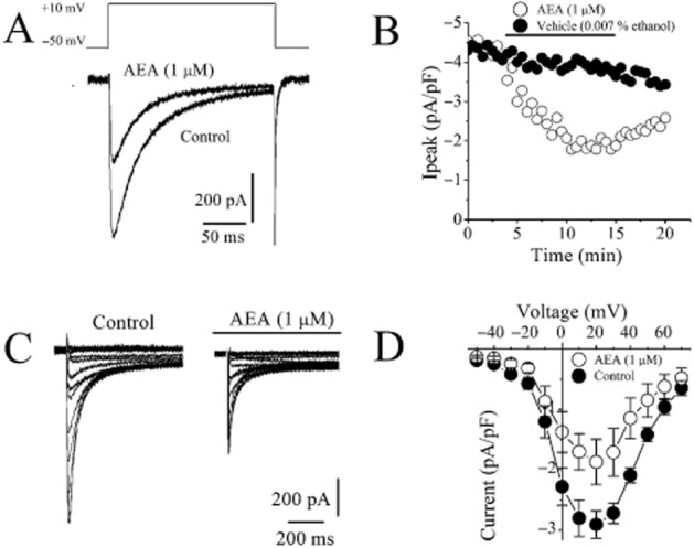
Figure 4.

Effect of AEA on Ca2+ currents mediated by L-type Ca2+ channels in rat ventricular myocytes. (A) AEA inhibits L-type Ca2+ currents recorded using whole-cell voltage-clamp mode of patch-clamp technique. Current traces recorded before (control) and after 10 min application of 1 μM AEA. ICa were recorded during 300 ms voltage pulses to +10 mV from a holding potential of −50 mV. (B) Averages of the maximal currents of VGCCs presented as a function of time in the presence of vehicle and 1 μM AEA; n = 5 cells. Application time for the agents is shown as a horizontal bar. (C) Representative recordings of ICa in response to the depicted pulse protocol under control conditions and after application of 1μM AEA. (D) Normalized and averaged I–V relationships of control ICa and ICa in the presence of 10 μM AEA determined by applying a series of step depolarizing pulses from −70 to +70 mV in 10 mV increments for a duration of 300 ms. Data shown are means ± SEM; n = 5–7 cells.
