Abstract
The case of a 77-year-old woman with symptoms of gastric outlet obstruction is presented. Transabdominal ultrasonography findings were suspicious of Bouveret's syndrome. Upper endoscopy confirmed this diagnosis. Bouveret's syndrome is a rare complication of gallstone disease caused by a bilioenteric fistula leading to gastric outlet obstruction by a gallstone and should be suspected in any patient who presents with pneumobilia without recent endoscopic retrograde cholangiopancreatography or biliary surgery.
Key words: Biliary diseases, Bilioenteric fistula, Bouveret's syndrome, Gallstones, Unspecific abdominal symptoms, Gastrointestinal obstruction, Pneumobilia
Introduction
Gallstone disease is very common in the United States and Western Europe with a prevalence of around 10%. However, 70–80% of patients remain asymptomatic [1]. Bouveret's syndrome – like Mirizzi syndrome – is a rare complication of gallstones. While Mirizzi syndrome means compression of the common bile duct by a jammed gallstone in the cystic duct leading to biliary obstruction, Bouveret's syndrome causes gastric outlet obstruction by a gallstone that enters the small bowel through a bilioenteric fistula and gets impacted in the duodenum or stomach [1, 2].
Case Report
A 77-year-old woman was admitted to the Accident and Emergency Department complaining of postprandial abdominal pain for the last 10 days. She had noted a single spike of fever (38.5°C) during the previous week and had been avoiding food because of postprandial abdominal pain. Her past medical history included only arterial hypertension. She denied any previous abdominal surgery. On physical examination vital signs were normal, the abdomen was soft and non-tender with normal bowel sounds. Complete blood count and electrolytes were normal. Liver enzymes [ASAT: 114 U/l (normal <40 U/l); ALAT: 131 U/l (normal <55 U/l)] and C-reactive protein [63 mg/l (normal <8 mg/l)] were elevated.
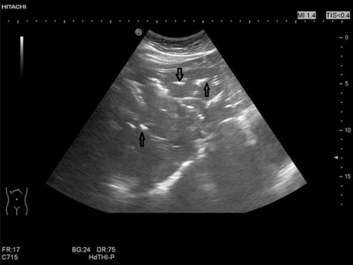
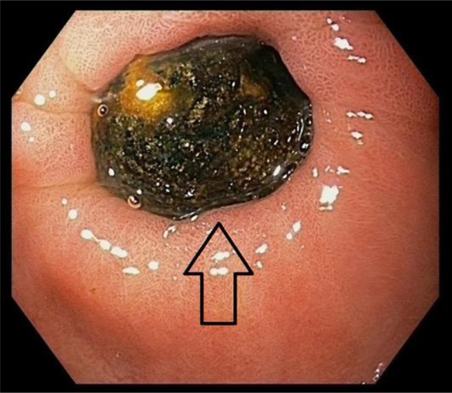
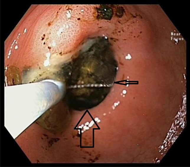
Transabdominal ultrasonography showed marked pneumobilia, a non-detectable gallbladder and dilation of the intrahepatic biliary tracts (fig. 1). Suspecting Bouveret's syndrome, we performed upper endoscopy and found an impacted gallstone in the duodenal bulb (fig. 2, see online suppl. videos 1 and 2; for all online suppl. material, see www.karger.com/doi/10.1159/000364818). Despite disproportion between the diameter of the stone and the pylorus, we tried to extract the gallstone endoscopically through the pylorus, but did not succeed (fig. 3, see online suppl. video 3). The case was then discussed with our surgeons. Open gastrotomy with stone extraction was performed. There were no postoperative complications.
Fig. 1.
Transabdominal ultrasonography showing marked pneumobilia. Pneumobilia means gas within the bile ducts (shown by arrows).
Fig. 2.
Endoscopy revealed an impacted gallstone (arrow) in the duodenal bulb, causing outlet obstruction in Bouveret's syndrome.
Fig. 3.
Endoscopic attempt to retrieve the stone, using a polypectomy snare (large arrow: stone; small arrow: snare).
Discussion
Bouveret's syndrome was first described in two case reports by Léon Bouveret in 1896 [3]. The current literature only reports single cases and few very small case series with up to 6 patients [4] with Bouveret's syndrome.
Only 0.3–5% of all patients with gallstones develop a bilioenteric fistula. Local perforation of the gallbladder wall can occur because of chronic inflammation and increased pressure to the gallbladder wall. This can cause local necrosis and lead to fistula formation and migration of a gallstone to the duodenum. Large gallstones measuring more than 2.5 cm can get impacted within the gastric outlet.
Bouveret's syndrome occurs primarily in elderly women, which is likely due to the higher incidence of gallstones in women. The bilioenteric fistula stays asymptomatic in 40–50% of cases until the gallstones pass from the gallbladder to the duodenum. Interestingly, the symptoms of gastric outlet obstruction are often unspecific, with epigastric pain, nausea, vomiting and subileus [5, 6, 7]. Sometimes fever, gastrointestinal bleeding and rarely icterus or signs of cholecystitis occur [8, 9].
One should consider Bouveret's syndrome in patients with pneumobilia, without a history of biliary surgery or recent endoscopic retrograde cholangiopancreatography and with unspecific obstructive symptoms. Pneumobilia is present in nearly half of the cases, especially when the gallbladder is not well detectable.
Plain abdominal radiography may show the typical triad of pneumobilia, small bowel ileus and an X-ray-positive ectopic stone that is pathognomonic for Bouveret's syndrome. This so-called Rigler's triad was first described by Leo G. Rigler [5, 10]. Even though this triad is highly specific for Bouveret's syndrome, it is only present in 20% of all cases. Computed tomography is considered the best diagnostic method [1, 4].
Endoscopy is the first-line treatment approach, in particular because of old age and comorbidities in these patients and the greater perioperative risk of surgical treatment. However, endoscopic treatment has a success rate of only 10%. If unsuccessful as in our case, patients have to undergo surgery for stone extraction [1].
Conclusion
Bouveret's syndrome should be suspected in any patient who presents with pneumobilia without recent endoscopic retrograde cholangiopancreatography or abdominal biliary surgery. Obstructive symptoms in elderly women are typically only mild and often unspecific.
Disclosure Statement
No conflicts of interests are declared by the authors.
Supplementary Material
Supplemental Video
Supplemental Video
Supplemental Video
References
- 1.Koulaouzidis A, Moschos J. Bouveret's syndrome. Narrative review. Ann Hepatol. 2007;6:89–91. [PubMed] [Google Scholar]
- 2.Langhost J, Schumacher B, Deselaers T, Neuhaus H. Successful endoscopic therapy of a gastric outlet obstruction due to a gallstone with intracorporeal laser lithotripsy: a case of Bouveret's syndrome. Gastrointest Endosc. 2000;51:209–213. doi: 10.1016/s0016-5107(00)70421-4. [DOI] [PubMed] [Google Scholar]
- 3.Bouveret L. Sténose du pylore adhérent à la vésicule. Rev Med (Paris) 1896;16:1–16. [Google Scholar]
- 4.Cappell MS, Davis M. Characterization of Bouveret's syndrome: a comprehensive review of 128 cases. Am J Gastroenterol. 2006;101:2139–2146. doi: 10.1111/j.1572-0241.2006.00645.x. [DOI] [PubMed] [Google Scholar]
- 5.Nickel F, Müller-Eschner MM, Chu J, von Tengg-Kobligk H, Müller-Stich BP. Bouveret's syndrome: presentation of two cases with review of the literature and development of a surgical treatment strategy. BMC Surg. 2013;13:33. doi: 10.1186/1471-2482-13-33. [DOI] [PMC free article] [PubMed] [Google Scholar]
- 6.Singh AK, Shirkhoda A, Lal N, Sagar P. Bouveret's syndrome: appearance on CT and upper gastrointestinal radiography before and after stone obturation. AJR Am J Roentgenol. 2003;181:828–830. doi: 10.2214/ajr.181.3.1810828. [DOI] [PubMed] [Google Scholar]
- 7.Meyenberger C, Michel C, Metzger U, Koelz HR. Gallstone ileus treated by extracorporeal shockwave lithotripsy. Gastrointest Endosc. 1996;43:508–511. doi: 10.1016/s0016-5107(96)70297-3. [DOI] [PubMed] [Google Scholar]
- 8.Clavien PA, Richon J, Burgan S, Rohner A. Gallstone ileus. Br J Surg. 1990;77:737–742. doi: 10.1002/bjs.1800770707. [DOI] [PubMed] [Google Scholar]
- 9.Moss JF, Bloom AD, Mesleh GF, Deziel D, Hopkins WM. Gallstone ileus. Am Surg. 1987;53:424–428. [PubMed] [Google Scholar]
- 10.Lewicki A. The Rigler sign and Leo G. Rigler. Radiology. 2004;233:7–12. doi: 10.1148/radiol.2331031985. [DOI] [PubMed] [Google Scholar]
Associated Data
This section collects any data citations, data availability statements, or supplementary materials included in this article.
Supplementary Materials
Supplemental Video
Supplemental Video
Supplemental Video


