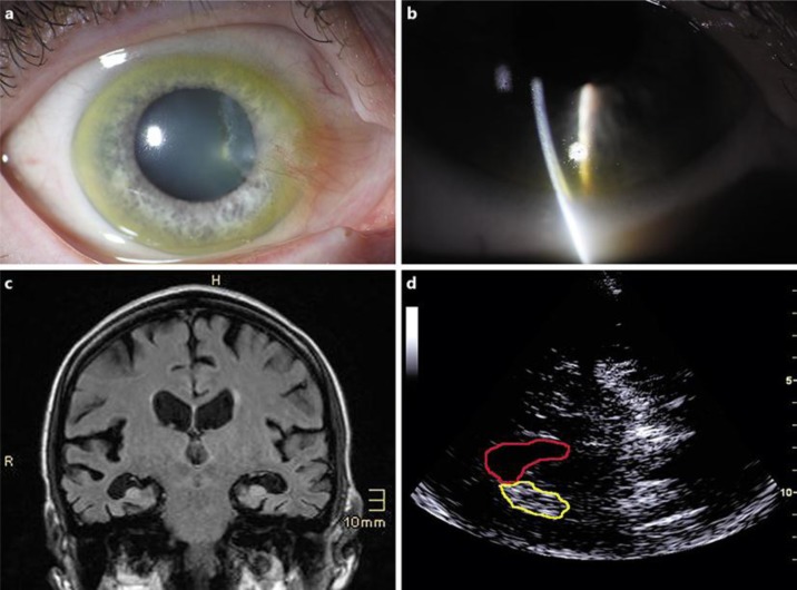
Fig. 1.
Ophthalmological findings: macroscopic view (a) and slit-lamp examination (b) revealed copper depositions within the Descemet membrane. c Coronary T1-weighted MRI showed temporoparietal-accentuated brain atrophy. d Sonography of the basal ganglia revealed hyperechogenicity of both lentiform nuclei, demonstrated here for the right lentiform nucleus (yellow) neighboring the right lateral ventricle (red).

