
Figure 6.
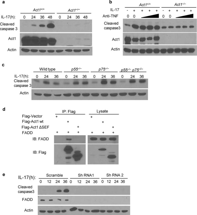
IL-17 induces cell apoptosis in an Act1 and FADD-dependent pathway. (a) Immunoassay of Act1+/+ or Act1−/− MEFs treated with IL-17 (50ng/ml) for the indicated times. Cells lysates were ran on SDS-PAGE gel and then probed for cleavaged caspase 3, Act1 and β-Actin. (b) MEFs treated with IL-17(50ng/ml) plus increasing amounts of anti-TNF-α neutralizing antibodies (1, 5, 10μg/ml). (c) Immunoblot analysis of wild type, p55-deficient, p75-deficient, p55 and p75 double-deficient kidney epithelial cells treated with IL-17 (200ng/ml) for the indicated times. (d) HEK293 cells were transfected with Flag-tagged vector as control, Flag -Act1 and – FADD. Lysates were immunoprecipitated with anti-Flag. (e) Immunoassay of OPCs transduced with lentivirus-expressing scrambled shRNA, or FADD-targeting shRNA1 or shRNA2. Cells were untreated or treated with IL-17 (200ng/ml). Cell lysates were blotted as indicated. Data are representative of at least three independent experiments. Full-length blots are presented in Supplementary Figure 6.