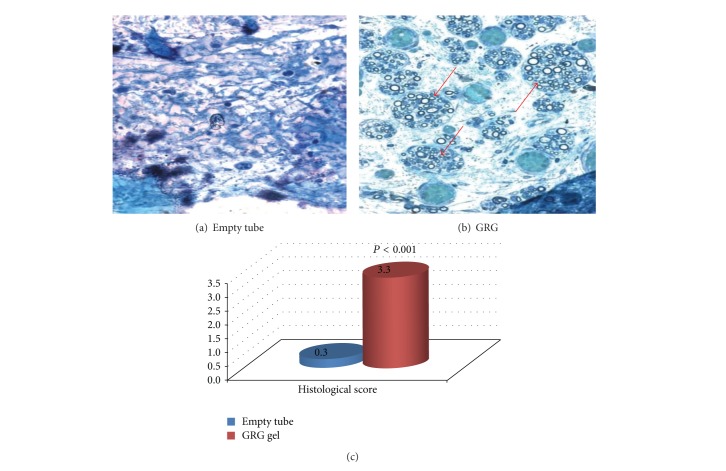
Figure 2.
(a) No axons, connective scar tissue; (b) massive growth of regenerative axons into the tube. The graph reflects histological score of the distal part of the nerve (blind examination) in difference between amounts of axons in the GRG group (good amount of axons with tendency to large-diameter axons) versus empty tube (scar tissue—no axons).

