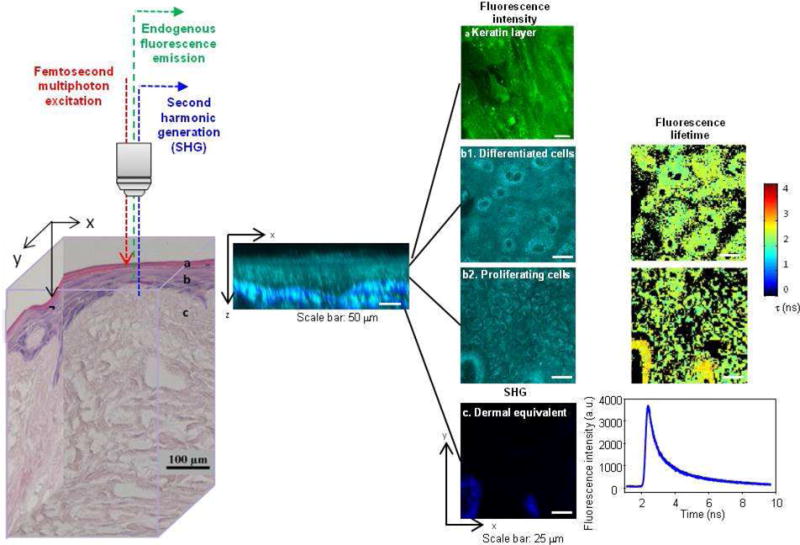
Figure 1.

Label-free nonlinear optical molecular imaging non-invasively interrogated a living EVPOME construct in three dimensions, measuring cross-sectional (middle) and en-face (right) images. EVPOME’s layered structure is evident in the histology (left) and in the cross-sectional image (middle) of cellular autofluorescence (NAD(P)H, cyan) with overlaid scaffold SHG (collagen, blue). EVPOMEs were composed of (1) a stratified cellular layer with > 3 cellular layers, including proliferative basal cells and differentiating cells, (2) attachment of basal cells to the dermal equivalent, and (3) a well-defined keratin layer. These three criteria were employed later for histological evaluation of construct viability. Fluorescence intensity, fluorescence lifetime, and SHG microscopic images were acquired to quantitatively assess EVPOME viability (right). Three-dimensional spatially-localized optical measurements interrogated cellular metabolic function and spatial organization (steady-state imaging) as well as cellular microenvironment (FLIM).
