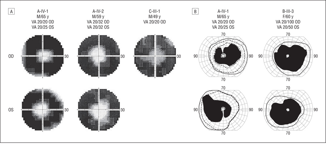
Figure 3.
Restricted visual fields in the central 60° among patients with KLHL7 mutations. A, Three patients (age range, 49–65 years) showed restricted visual fields to central 10° to 20° using the Humphrey visual field analyzer protocol (30-2 SITA Fast Program; Humphrey Instruments, San Leandro, California). High-density regions represent lost visual fields; gray regions indicate transitional zones. B, Goldmann perimetry shows visual field retention in the far periphery (solid lines indicates V-4-e; dashed lines, III-4-e). VA indicates visual acuity.

