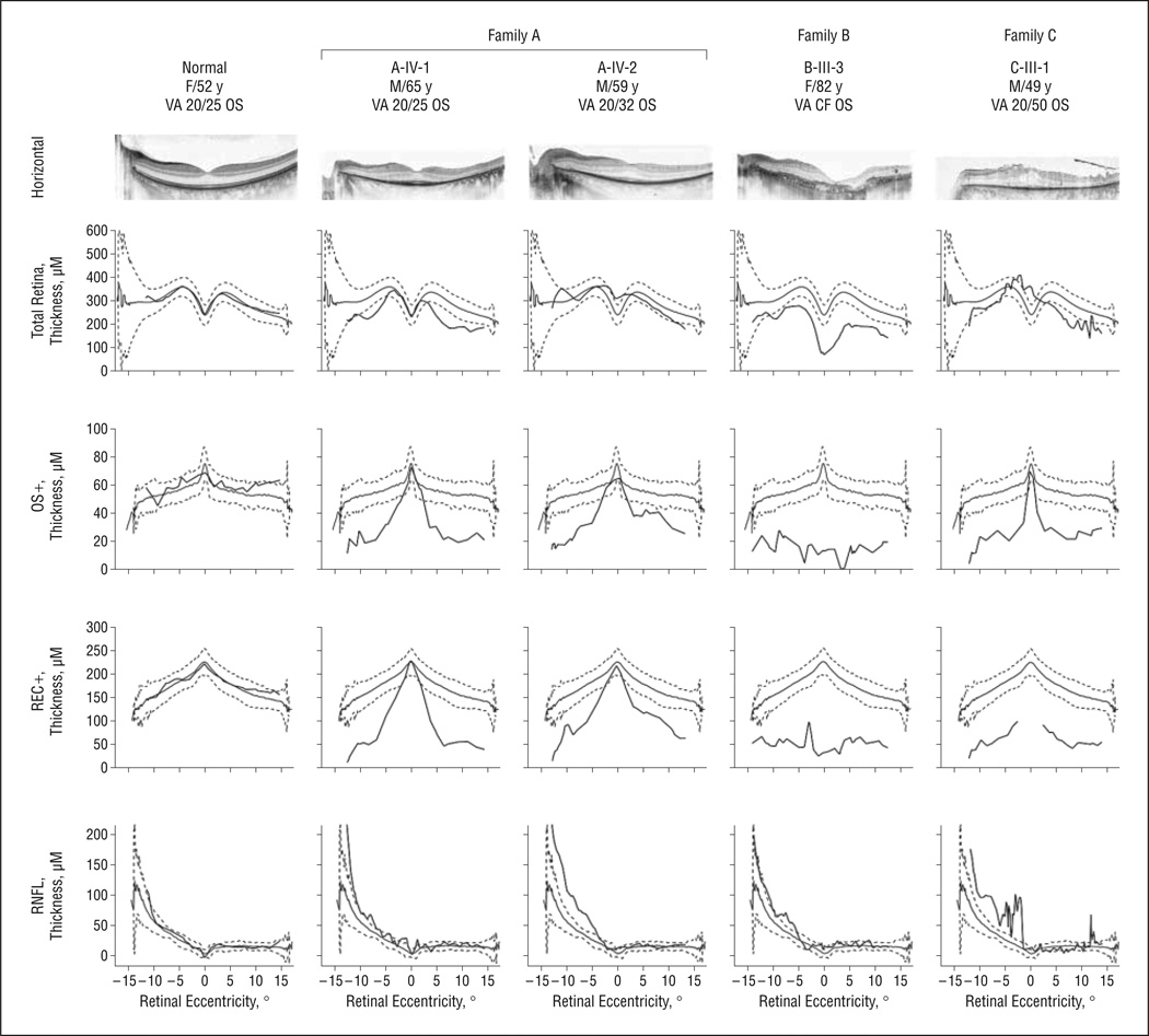
Figure 6.
Spectral-domain optical coherence tomography images acquired from patients carrying KLHL7 mutations (central 30° and horizontal meridian). Images were segmented to show the thickness profile of retinal nerve fiber layer (RNFL), total retina (neural retina plus retinal pigment epithelium), OS+ (photoreceptor outer segment and retinal pigment epithelium), and REC+ (retinal pigment epithelium, photoreceptor outer segment, inner segment, outer nuclear layer, and outer plexiform layer). CF indicates counting fingers; VA, visual acuity.

