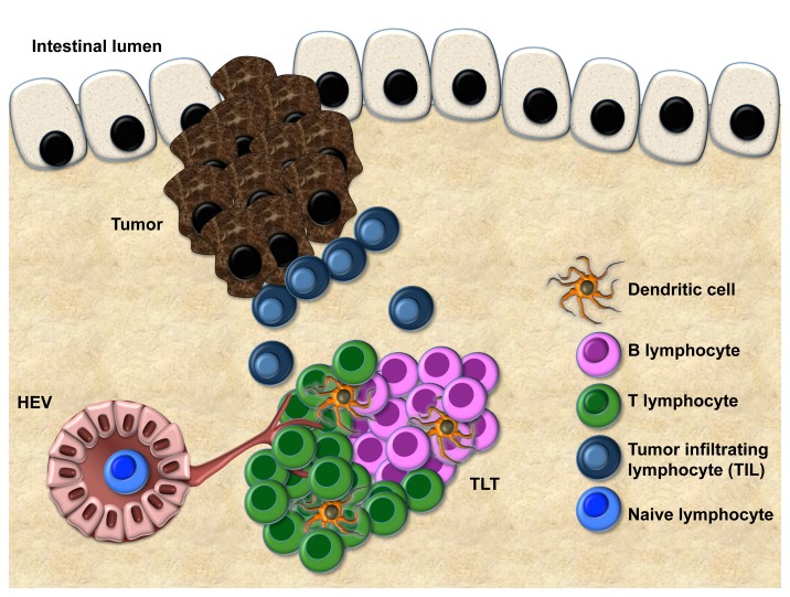
Figure 1. Features of tertiary lymphoid tissue (TLT) at the tumor margin. Compartmentalized areas of T cells and B cells, dendritic cells, and a vascular network, including lymphatic vessels and high endothelial venules (HEV) are shown. The correlation between the extent of tertiary lymphoid tissue (TLT) and T-cell infiltration suggests that TLT represents a gateway for tumor-infiltrating T cells (TILs).

An official website of the United States government
Here's how you know
Official websites use .gov
A
.gov website belongs to an official
government organization in the United States.
Secure .gov websites use HTTPS
A lock (
) or https:// means you've safely
connected to the .gov website. Share sensitive
information only on official, secure websites.
