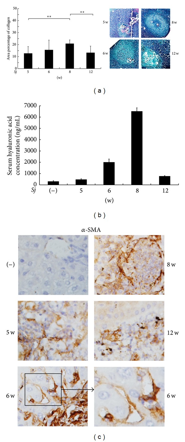
Figure 1.

Extent of mouse hepatic fibrosis increased gradually after Sj infection. BALB/c mice were infected with 20 ± 3 infective cercariae of Sj for 5, 6, 8, and 12 weeks, and noninfected mice served as negative control. Liver tissues were fixed and stained with Masson trichrome or anti-α-SMA antibody (original magnification 40x). Collagen deposition area percentage (Masson trichrome staining positive area percentage of each section) was calculated and shown in (a, left panel). Representative liver granulomas stained with Masson's trichrome staining are shown in (a, right panel) at 20x magnification. Concentration of hyaluronic acid in mouse liver serum is shown in (b) through ELISA assay. Expression level of α-SMA protein in mouse liver tissues is shown in (c) through IHC assay. Data are presented as mean ± SD from eight mice per group. Experiments were performed twice. **P < 0.01.
