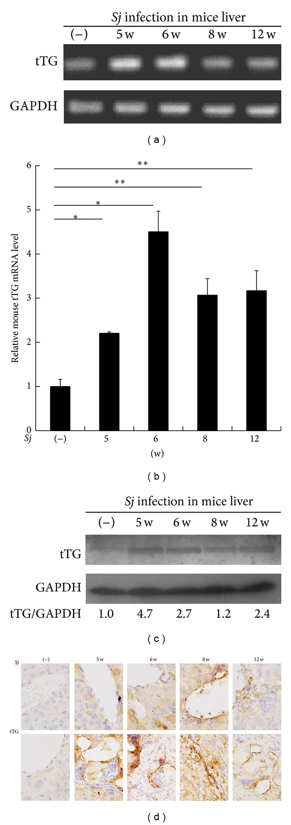
Figure 3.

tTG expression was upregulated progressively in mice liver after Sj infection. (a, b) tTG mRNA level in BABL/c mice liver is upregulated at weeks 5, 6, 8, and 12 after Sj infection, as determined through RT-PCR and Q-PCR. GAPDH is detected as an internal control. Data are presented as mean ± SD from eight mice per group. *P < 0.05 and **P < 0.01 compared with noninfected mice (−). (c) tTG protein level of mice liver homogenates is tested by Western blot analysis. GAPDH is used as loading control. (d) Different localization expression levels of tTG protein in BALB/c mice liver are shown through IHC assay at weeks 5, 6, 8, and 12 after Sj infection. Upper panel: extent of tTG positive staining in hepatic cell and liver tissue around the liver sinusoid. Lower panel: extent of tTG positive staining in the granuloma and nearby liver tissue after Sj egg deposition. Representative tTG staining sections are shown at 40x magnification.
