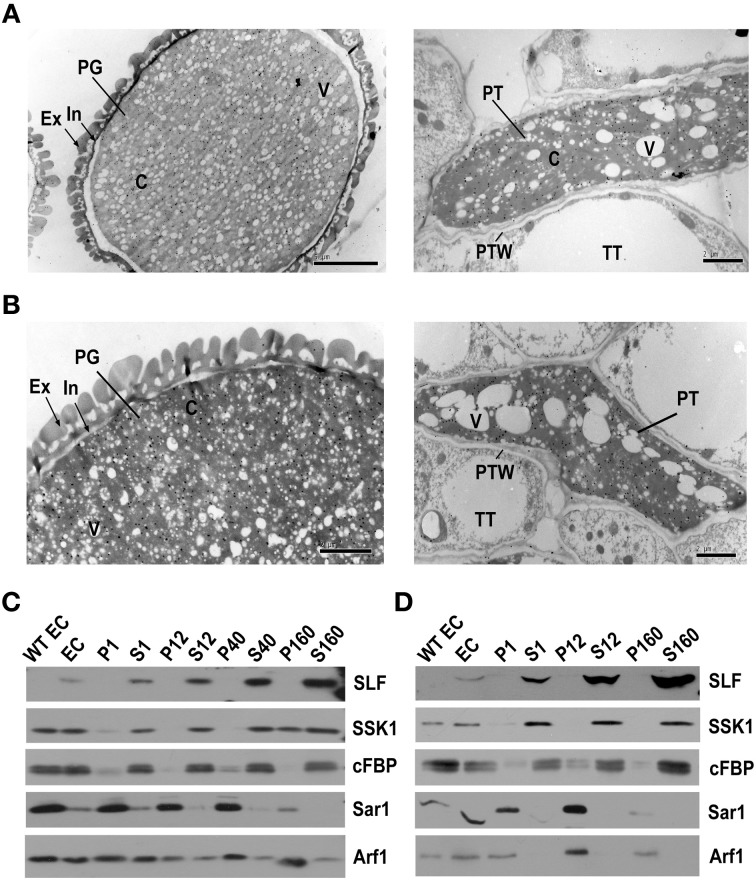
Figure 1.
PhS3L-SLF1 and PhSSK1 are both located in the cytosol of pollen grains and pollen tubes. (A) Immunogold labeling of PhS3L-SLF1::FLAG in pollen grain (left) and pollen tube (right). (B) Immunogold labeling of PhSSK1 in pollen grain (left) and pollen tube (right). PG, pollen grain; Ex, exine; In, intine (arrow); C, cytosol; V, vacuole; PT, pollen tube; PTW, pollen tube wall; TT, transmitting tract tissue. (C) Western blot detection of PhS3L-SLF1::FLAG (SLF) and PhSSK1 (SSK1) in subcellular fractions of pollen grains. (D) Western blot detection of PhS3L-SLF1::FLAG (SLF) and PhSSK1 (SSK1) in subcellular fractions of in vitro germinated pollen tubes. WT EC and EC denote entire cell homogenates from wild-type and the transgenic pollen grains or pollen tubes, respectively. Pellet fractions (P1, P12, P40, and P160) and supernatant fractions (S1, S12, S40, and S160) are derived from differential centrifugation at 1000, 12,000, 40,000, and 160,000 g, respectively. cFBP, Sar1, and Arf1 are marker antibodies for cytosol, endoplasmic reticulum (ER) and Golgi, respectively.

