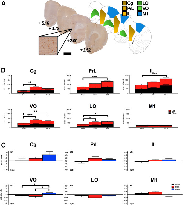
Figure 3.

c-fos expression in the prefrontal cortex. (A) Exemplificative c-fos stained sections, the respective Paxinos and Watson atlas diagrams [7] and respective distance to Bregma (mm) are given. Scale bar, 2 mm (100 μm inset). (B) SNI groups present a higher c-fos density when compared to sham controls in the mPFC and OFC but not in the control area M1. The hemisphere side was not a determining factor for the differences between the groups. (C) SNI-R contrary to sham/SNI-L presented a leftward shift in the laterality index in the VO following ASST completion. The functional integrity of this area is critical to Rev execution. *P < 0.05, **P < 0.01 and ***P < 0.001. Data presented as mean ± S.E.M.
