Figure 3.

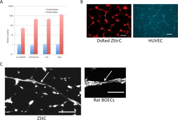
ZStrCs can be induced to express endothelial specific genes and are able to form capillary networks and endotubes on matrigel. (A) qRT-PCR of endothelial-specific genes expressed by ZStrCs in normal growth media versus EGM2 media. (B) Top panel shows zebrafish stromal cells isolated from a transgenic zebrafish constitutively expressing DsRed that were plated onto matrigel-precoated 96-well plates in EGM-2 media and incubated at 32°C for 24 hours followed by fluorescent microscopy. Shown is a typical capillary network. Scale bar indicates 100 microns. Bottom panel shows a similar endothelial differentiation using HUVECs labeled with CellTracker™Violet. Scale bar indicates 200 microns. (C) An example of a lumen-containing endotube (white arrow) formed by ZStrC or rat BOECs on matrigel. Confocal images were acquired using an Olympus Fluo View FV 1000 Inverted microscope in gray scale mode and final image generated as a projection of 27 image planes. Scale bar indicates 100 microns.
