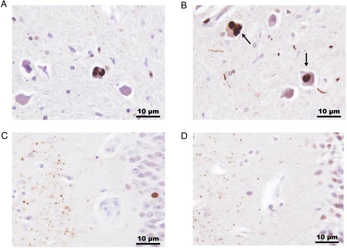
Figure 2.
Immunopositive ubiquilin-2 inclusions in brain stem and hippocampus from two affected members of family 1T. Ubiquilin-2 positive intracytoplasmic aggregates (arrows) were identified in neurons in the inferior olivary nucleus along with sparse immunopositive neurites (A, individual II-3; B, individual III-3). Hippocampal aggregates of ubiquilin-2 were detected in the molecular layer (left side of section) of the fascia dentata (C, individual II-3; D, individual III-3). Ubiquilin-2 immunohistochemistry was performed with an ubiquilin-2 polyclonal antibody (Sigma Life Sciences) using diamobenzidine and a hematoxylin counterstain, x400 original magnification.

