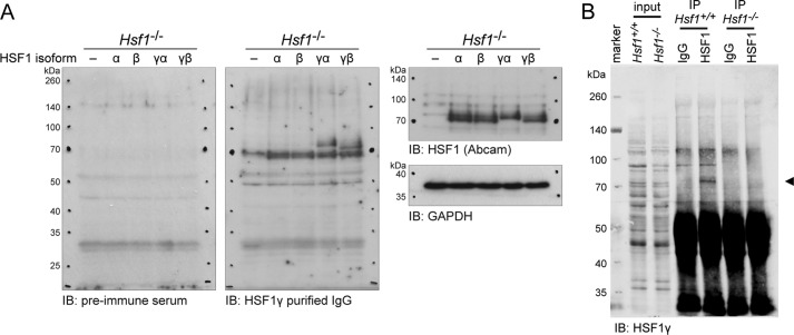
FIGURE 3.
The Hsf1γ isoforms are translated into protein in vivo. A, verification of the anti-HSF1γ antibody raised against the exon 9a peptide. An Hsf1 knock-out cell line (Hsf1−/−) was transfected with either vector (−) or the four HSF1 isoforms (α, β, γα, γβ). Proteins were extracted and separated by SDS-PAGE. The membrane was first incubated with HSF1γ preimmune serum. After image acquisition, the membrane was stripped and incubated with anti-HSF1γ purified IgG. Thirdly, the blot was stripped again, cut, and incubated with anti-HSF1 (Abcam), or GAPDH, respectively. IB = immunoblot detection antibody. B, IP of HSF1 from mouse testes. Testes from wild type (Hsf1+/+) and knock-out (Hsf1−/−) mice were homogenized, and HSF1 was immunoprecipitated as described under “Experimental Procedures.” Following Western blotting, the membrane was cut at about 60 kDa, but each part was incubated with identical conditions and reassembled for image acquisition. HSF1γ isoforms were detected with anti-HSF1γ purified IgG (◀) in the IP from wild type, but not from knock-out mice.

