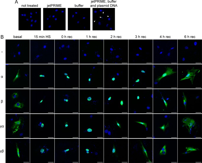
FIGURE 5.
HSF1 isoforms show different nuclear export or degradation kinetics after heat shock. A, DAPI stain (blue) of Hsf1 knock-out cells with different transfection conditions. Cells were completely untreated (not treated panel), transfected with only the jetPRIME reagent (jetPRIME panel) or only with the buffer (buffer panel), or transfected with both together with plasmid DNA (jetPRIME, buffer and plasmid DNA panel). The white arrows indicate the appearance of DAPI-positive small puncta when DNA is transfected. B, immunofluorescence of HSF1 isoforms after heat shock (HS). Cells were either kept at 37 °C (basal panel) or heat-shocked for 45 min at 42 °C and recovered for the indicated time period (0–6 h rec) at 37 °C. The second column shows cells that were only heat-shocked for 15 min (15 min HS panel) at 42 °C and immediately fixed. Images are shown as merged color with DAPI in blue and anti-FLAG (FLAG-tagged HSF1 isoforms) in green. White bars correspond to 50 μm. rec = recovery.

