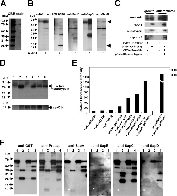
FIGURE 2.
Effect of epidermal proteases on prosaposin processing. A, Coomassie Brilliant Blue staining of GST-prosaposin. Recombinant GST-prosaposin was subjected to SDS-PAGE and stained with Coomassie Brilliant Blue. The band of the recombinant protein can be seen at ∼75 kDa. B, Western blot analysis of prosaposin degradation products. Extracts from differentiated keratinocytes containing prosaposin were incubated with revC14. Western blot analysis were carried out using antibodies to prosaposin (anti-Prosap), saposin A (anti-SapA), sapoins B (anti-SapB), saposin C (anti-SapC), and saposin D (anti-SapD). During incubation with active caspase-14, prosaposin in the extract was degraded into multiple intermediate products. The glycosylated form of saposin A (15 kDa) was detected. Arrowhead, GST-prosaposin; arrow, saposin A. C, co-transfection of pCMV-HA-Prosap, pCMV-HA-revC14, and pCMV-HA-mesotrypsin in growth and differentiated phases. The Western blot was carried out using a specific antibody to each molecule. D, detection of the active form of mesotrypsin in revC14-transfected keratinocytes. Keratinocytes were transfected with pCMV-HA-vector (control), pCMV-HA-revC14, or pCMV-HA-mesotrypsin and further incubated for 24 h in the presence or absence of protease inhibitors. Cell extracts were subjected to SDS-polyacrylamide gel electrophoresis and the presence of mesotrypsin was analyzed by Western blotting using anti-mesotrypsin antibody. Lane 1, pCMV-HA-vector; lane 2, pCMV-HA-revC14; lane 3, pCMV-HA-revC14 + Z-VAD-fmk; lane 4, pCMV-HA-revC14 + leupeptin; lane 5, pCMV-HA-revC14 + Z-VAD-fmk + leupeptin; lane 6, pCMV-HA-mesotrypsin. E, effect of caspase-14 on mesotrypsinogen activation. Enzymatic activity of mesotrypsin was measured using Boc-Gln-Ala-Arg-methylcoumarin amide as a substrate after incubation with caspase-14. To evaluate the direct hydrolytic activity of caspase-14 on this substrate, the same concentration of caspase-14 was incubated without mesotrypsinogen. Amounts of enzymes used in each assay (ng) are listed in parentheses. For comparison, enterokinase was also used. Results are shown as the mean of duplicate experiments. F, Western blot analysis of prosaposin degradation products by mesotrypsin, KLK5, and KLK7. After incubation with each protease, prosaposin degradation products were detected with antibodies to GST, prosaposin, saposin A, saposin B, saposin C, and saposin D. Asterisks indicate the presence of each saposin protein band. Lane 1, prosaposin control; lane 2, prosaposin + mesotrypsin; lane 3, prosaposin + KLK5; lane 4, prosaposin + KLK7.

