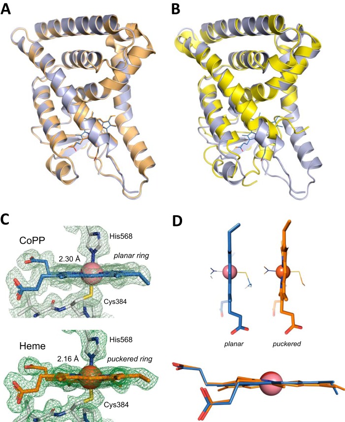
FIGURE 3.
Crystal structure of CoPP bound to the REV-ERBβ-LBD. A, overlay of CoPP- (blue) and heme- (orange; PDB code 3CQV) bound structures of the REV-ERBβ-LBD. B, overlay of the CoPP-bound (blue) and apo (yellow; PDB 2V0V) structures of the REV-ERBβ-LBD. C, cobalt-His-568 coordination distance (2.30 Å). The distance is 0.14 Å larger than the one observed for the iron-His-568 holo structure (2.16 Å) (PDB code 3CQV (11)). D, CoPP adopts a planar conformation in the LBD of REV-ERBβ-LBD different from the non-planar, puckered distortion observed for the heme group.

