FIGURE 3.
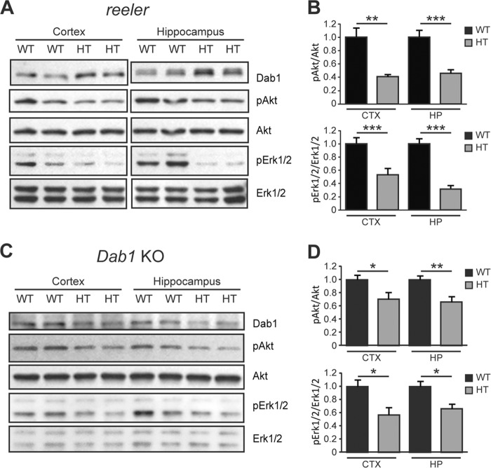
Abnormal Akt and Erk1/2 signaling in juvenile heterozygous reeler and Dab1 KO mice. A, Western blot analysis of forebrain regions from 3–4-week-old wild type (WT) and heterozygous (HT) reeler mice. The levels of phospho-Akt and phospho-Erk1/2 were significantly reduced both the cerebral cortex and hippocampus of reeler mice. B, data were quantified from n = 9 WT and n = 7 HT mice of the reeler strain. C, Western blot analysis of cortex and hippocampus from 3–4-week-old WT and HT Dab1 KO mice. The phosphorylation levels of Akt and Erk1/2 were decreased significantly in HT mice. D, data were quantified from n = 10 WT, n = 7 HT mice of the Dab1 KO strain.

