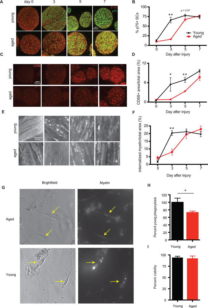
Figure 7. Functional defects in aged Schwann cells.
Cross-sections and whole mounts of sciatic nerves from young and aged animals at day 0, 3, 5 and 7 after nerve crush injury stained with (A) S100 and p75, (B) CD68, and (C) MBP. (D, E and F) Quantification of staining from (A, B and C). Mean ± SEM is plotted, n = 3–4 mice per group. P < 0.05 by two-way ANOVA for (D–F). *P < 0.05, **P < 0.01 with mutliple t-tests, Holm-Sidak correction. SCs were purified from the sciatic nerves of young and aged animals 3 days after sciatic nerve crush injury and incubated with myelin for 2 hours. (G) Representative images of purified young and aged SCs with myelin (yellow arrows). (H) Quantification of myelin contained within SCs by FACS analysis. (I) Cell viability as determined by DAPI staining. n=3–4 per group. **P <0.01 with unpaired t-test.

