Fig. 6.

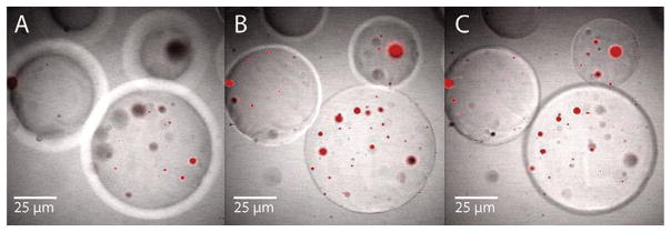
Confocal microscopy pictures of hybrid particles of Rhodamine-loaded PLGA MS incorporated in PEG MP. The micrographs are at optical depths of (A) 100 μm, (B) 350 μm and (C) 450 μm from the surface of the PEG MP.

Confocal microscopy pictures of hybrid particles of Rhodamine-loaded PLGA MS incorporated in PEG MP. The micrographs are at optical depths of (A) 100 μm, (B) 350 μm and (C) 450 μm from the surface of the PEG MP.