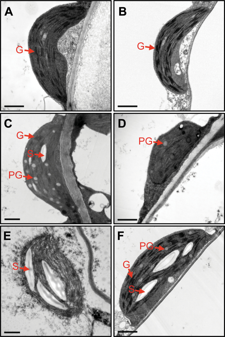
Fig. 3.
Transmission electron microscopy of plastids in atg5 leaves under mild salt-stress conditions. (A, B) Chloroplasts in the mesophyll cells of 3-week-old wild-type (WT; A) and atg5 (B) leaves before salt (150mM) treatment. (C–F) Chloroplasts in the mesophyll cells of WT (C, D, E) and atg5 (F) leaves after 4 DST. DST, days of salt treatment; G, grana thylakoid; PG, plastoglobule; S, starch. Scale bars=1 μm. (This figure is available in colour at JXB online.)

