
Figure 2.
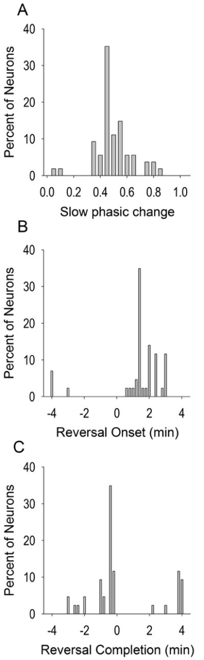
Slow phasic properties. The magnitude and direction of standardized slow phasic changes in firing (A) were fairly normally distributed. Most neurons exhibited reversal onsets (B) within the first two min and completions (C) occurred typically within two minutes prior to the infusion. Reversal onset and completion are expressed as time from the infusion (min). Note that for assessing reversal onset and completion, each spline curve (solid lines of Figure 1, see methods) was examined first from the time of the overall min (for decrease patterns) or overall max (for increase patterns) to the +4 min time bin. If reversal onset or completion was not observed during this epoch, the spline curve was further examined from the -4 min time bin to the time of the overall max (for decrease patterns) or overall min (for increase patterns). Figure 2A includes no change neurons while 2B-2C include only slow phasic neurons.