Figure 2.
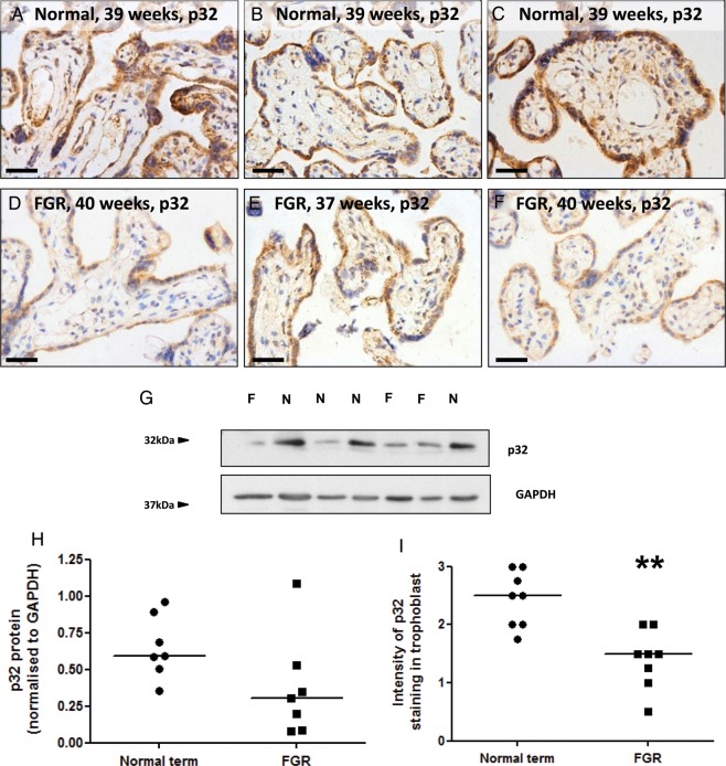
Trophoblast p32 expression is reduced in placentas from women with FGR. (A–F) Immunostaining of (A–C) normal term placentas or (D–F) placentas from women with FGR with an antibody to p32 (DAB labelling, brown). Nuclei were counterstained with haematoxylin (blue). Scale bar represents 50 µm. Images are representative of n = 8 normal and eight FGR samples. (G) Total p32 expression in normal term placentas (n = 7) or placentas from women with FGR (n = 7) was analysed by western blotting. (H) Expression was quantified by densitometry and normalized to GAPDH expression. Median, n = 7. (I) Trophoblast p32 staining intensity was quantified by two independent assessors. Median, n = 8; **P < 0.01, Mann–Whitney U-test.

