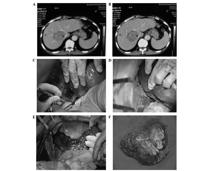
Figure 1.
Hepatectomy of segments IV, V, VIII and caudate lobe with cholecystectomy. (A and B) Computed tomography revealed a centrally located hepatocellular carcinoma invading the caudate lobe. (C) A long suture was placed behind the right hepatic vein. (D) The trunk of the left and middle hepatic veins was exposed and the middle hepatic vein was ligated. (E) Postoperative view of the liver. (F) Excised tumor specimen.

