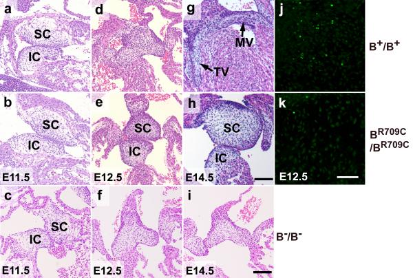
Figure 4.
Defects in Fusion and Remodeling of the Atrioventricular Cushions in BR709C/BR709C Mouse Hearts. a-i. H&E stained heart sections of B+/B+, BR709C/BR709C, and B−/B− embryos show developmental progression of atrioventricular (AV) cushions from E11.5 to E14.5. E11.5 AV cushions show no differences in size, morphology and positioning between B+/B+ (a), BR709C/BR709C (b) and B−/B− (c) hearts. B+/B+ AV cushions fuse and start to elongate at E12.5 (d), and acquire mature mitral (MV) and tricuspid (TV) valve leaflets by E14.5 (g). BR709C/BR709C cushions remain unfused and show no sign of maturation at E12.5 (e) and E14.5 (h). The fusion of AV cushions in B−/B− hearts appears normal at E12.5 (f), however further maturation into cardiac valves is delayed at E14.5 (i) compared to the B+/B+ mouse (e). IC, inferior AV cushion; SC, superior AV cushion. j,k. TUNEL assay shows defective apoptosis in developing BR709C/BR709C cushions. Apoptotic cells are readily seen in B+/B+ cushions (j, green), but very few apoptotic cells are found in BR709C/BR709C cushions (k). DAPI (blue) stains nuclei. Scale bar: a-i, 40 μm; j,k, 25μM.

