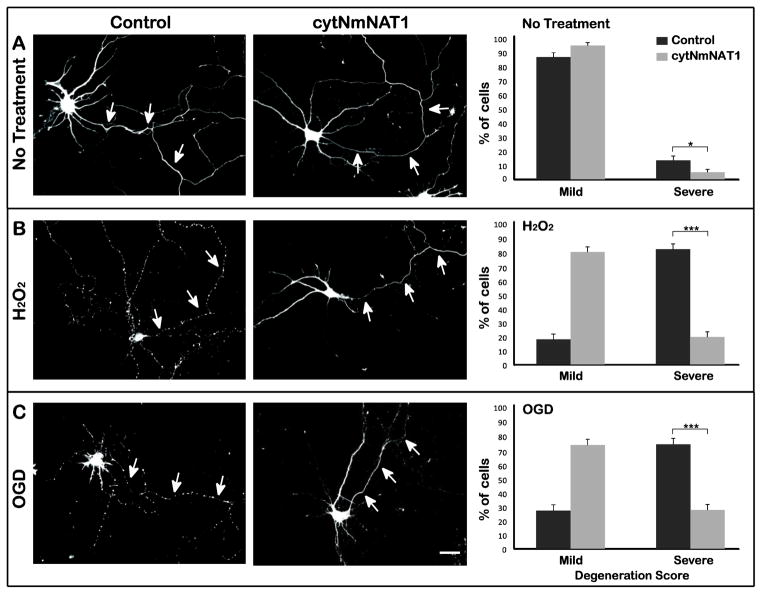
Figure 1. Expression of cytNmNAT1 protects the axons of cultured hippocampal neurons from hydrogen peroxide or OGD.
(A) Neurons were electroporated with either soluble GFP (control) or cytNmNAT1-GFP before plating. After 7–8 days in culture, neurons were challenged with either 100 μM hydrogen peroxide for 1 h (B) or oxygen glucose deprivation for 4 h (C). Axons from control neurons (i.e. expressing GFP alone) exhibited pronounced signs of degeneration following hydrogen peroxide exposure or OGD, as indicated by beading and fragmentation (B & C left column). In contrast, axons from neurons expressing cytNmNAT1-GFP were largely intact following the same challenges (B & C middle column). To quantify these results, the extent of degeneration was evaluated based on a scale ranging from 1 (minimal degeneration) to 4 (beading or fragmentation of the entire axonal arbor). Cells with a degeneration score of 1 or 2 were grouped together in a single category, referred to as “mild” degeneration, and those with scores of 3 and 4 were grouped together as “severe” degeneration. Exposure to hydrogen peroxide or OGD caused a large increase in the percentage of severely degenerated axons. In cells expressing cytNmNAT1-GFP, the percentage of severely degenerated axons was much reduced (B & C right column). Axonal degeneration was evaluated 6 h after hydrogen peroxide exposure and 24 h after OGD. * p < 0.05; *** P < 0.001. Scale bar: 30 μm.

