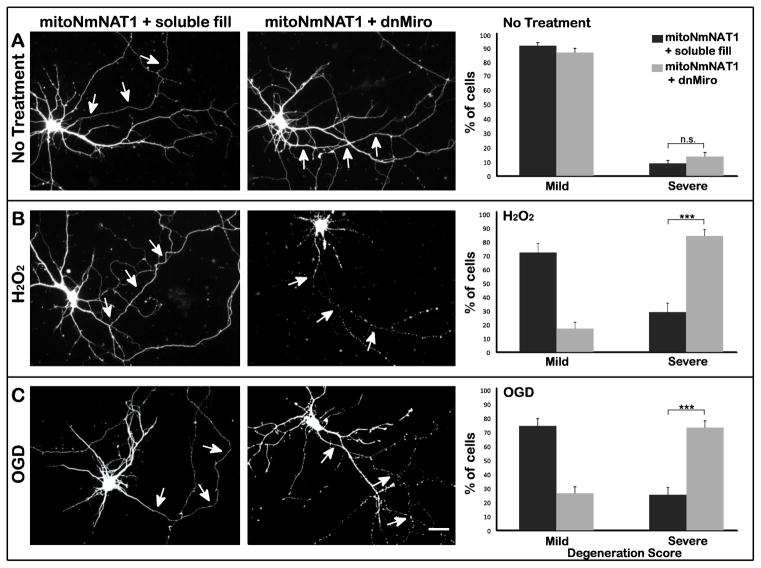
Figure 7. Inhibition of mitochondrial transport abolishes the axon-protective effects of mitochondrially targeted NmNAT1.
(A) Neurons were electroporated with mitoNmNAT1-GFP before plating, and then were transfected with either dnMiro-RFP or RFP alone as a control (soluble fill) at DIV 7. Neurons were subjected to hydrogen peroxide (B) or OGD (C) 20h after transfection. In neurons expressing mitoNmNAT1 and a soluble fill, axons showed little damage after either insult (B & C left column). In contrast, the axons of cells expressing dnMiro were badly fragmented (B & C middle column). Quantification confirmed that without injury, expressing dnMiro did not affect axonal integrity (P=0.2) (A right column). When treated with hydrogen peroxide, 83% of the axons were severely degenerated (scores of 3 or 4) in neurons expressing dnMiro, compared with only 28% in neurons expressing mitoNmNAT1 (B right column). After OGD, 73% of the axons of cells expressing dnMiro were severely degenerated, compared with 25% of axons in neurons expressing mitoNmNAT1 (C right column). *** p < 0.001. Scale bar=30 μm.

