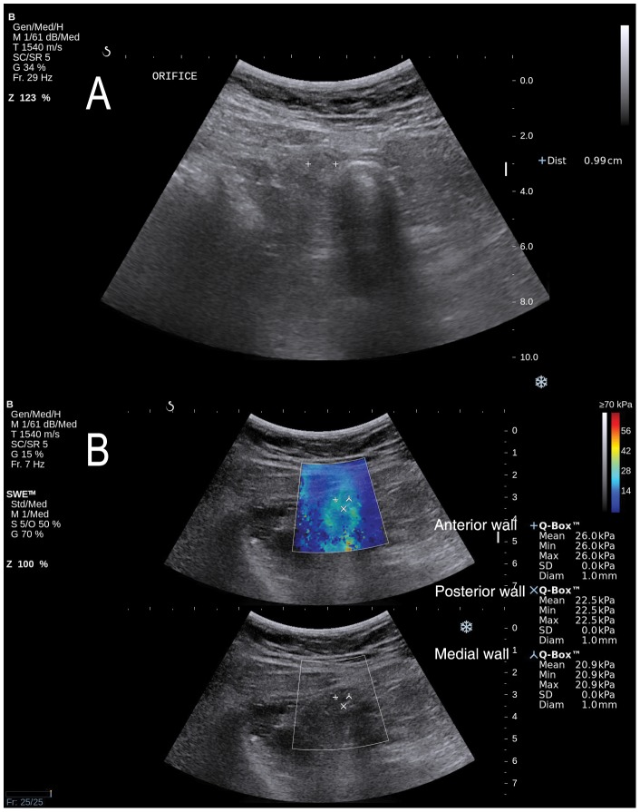
Figure 2. Gray-scale ultrasonography and shear wave elastography in a 47-year old female patient with appendicitis.
A. Gray-scale ultrasonography was 9.9 mm in diameter. The echogenicity of periappendiceal fat and the appendiceal wall thickening were also noted (not shown). B. Elastic modulus scales (mean Q-Box) by shear wave elastography were 26.0, 20.9, and 22.5 kilopascal (kPa) in the anterior, medial, and posterior wall of the appendix, respectively. The highest elastic modulus scale (26.0 kPa) was selected for analysis. The modified Alavarado score of this patient was 7, and computed tomography showed a larger diameter (≥6 mm), enhancement of periappendiceal fat and wall thickening. The histopathology result showed appendicitis.

