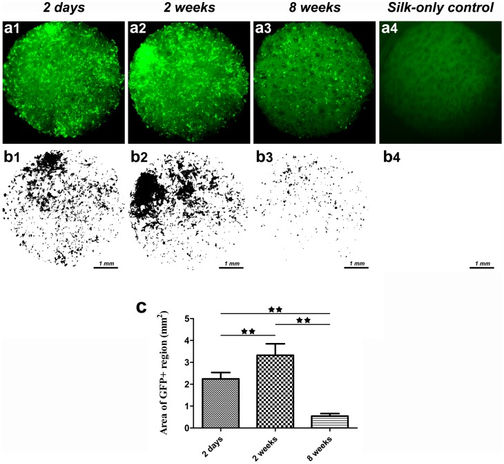
Figure 3. The fate of rat BMSCs cultured with silk scaffolds in vitro.
(a) Sequential observation of GFP labelled cells in the scaffolds by fluorescence microscopy. (b) Black-white images processed using Image J software. (c) The graph shows the area of GFP positive regions for the different study groups. (★★, represents p<0.01).

