
Figure 2. Gray matter volume reductions in the region-of interest (ROI) parahippocampal gyrus+hippocampus bilaterally.
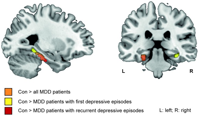
Gray matter volume reduction in ROI gyrus+hippocampus bilaterally in all MDD patients versus healthy controls (orange), patients with first depressive episode versus healthy controls (yellow) and patients with recurrent depressive episodes versus healthy controls (red) (Table 4). (Region-of-interest analyses, p<0.01, k = 109; view: MNI: −27 −29 −20).