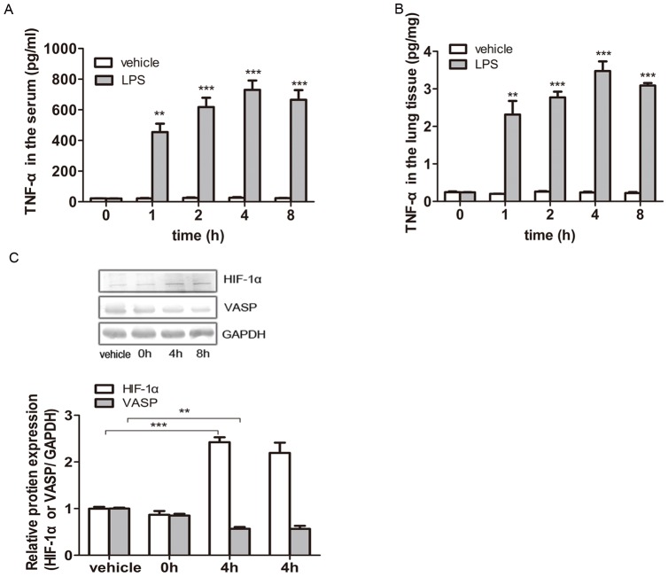
Figure 4. VASP was downregulated via the TNF-α-induced activation of HIF-1α during acute lung injury in vivo.
Balb/c mice were peritoneally injected with 0.01 mg/g LPS to model ALI or the same dose of saline in the vehicle group. The animals were treated for 0–8 hours. (A) After 0–8 hours of stimulation, the sera of the animal models were prepared via orbital blood collection, and ELISA was performed to detect the concentration of TNF-α in the serum. In the vehicle groups, the TNF-α concentration in the serum remained at a very low level (<30 pg/mL) and displayed no obvious changes over time (p>0.05 vs. vehicle). In contrast, the sera from mice treated with LPS for 0–8 hours all displayed much higher TNF-α concentrations (>450 pg/mL), which peaked at 4 hours (>700 pg/mL), compared with the vehicle groups (**p<0.01 and ***p<0.001 vs. vehicle). (B) After LPS treatment, the supernatant liquid of lung tissues was prepared from the excised left lungs, and ELISA was performed to detect the concentration of TNF-α in the lung tissues. The TNF-α concentration in the lung tissues also remained at a very low level (<0.3 pg/mg) in the vehicle groups and displayed no obvious changes over time (p>0.05 vs. vehicle). Conversely, the TNF-α concentration in the lung tissues from LPS-induced ALI mice was dramatically higher than that of the vehicle groups (>2.0 pg/mg; **p<0.01 and ***p<0.001 vs. vehicle) and peaked at 4 hours (>3.4 pg/mg). (C) The HIF-1α and VASP expression levels in mouse lung tissues were detected by western blotting. GAPDH served as a loading control. Representative blots from three independent experiments with similar results are shown. The relative protein expression levels obtained for HIF-1α or VASP/GAPDH are shown in the bar graphs. Compared with the vehicle group, the HIF-1α levels in the lung tissues were evidently elevated by 142.3% in the 4-hour group (***p<0.001) and 119.3% in the 8-hour group (**p<0.01 vs. vehicle). Correspondingly, VASP expression decreased by 43.1% in the 4-hour group (**p<0.01 vs. vehicle) and 43.5% in the 8-hour group (**p<0.01 vs. vehicle). The test was repeated three times with identical results. The data are presented as the mean ± SEM. *p<0.05, **p<0.01, ***p<0.001.

