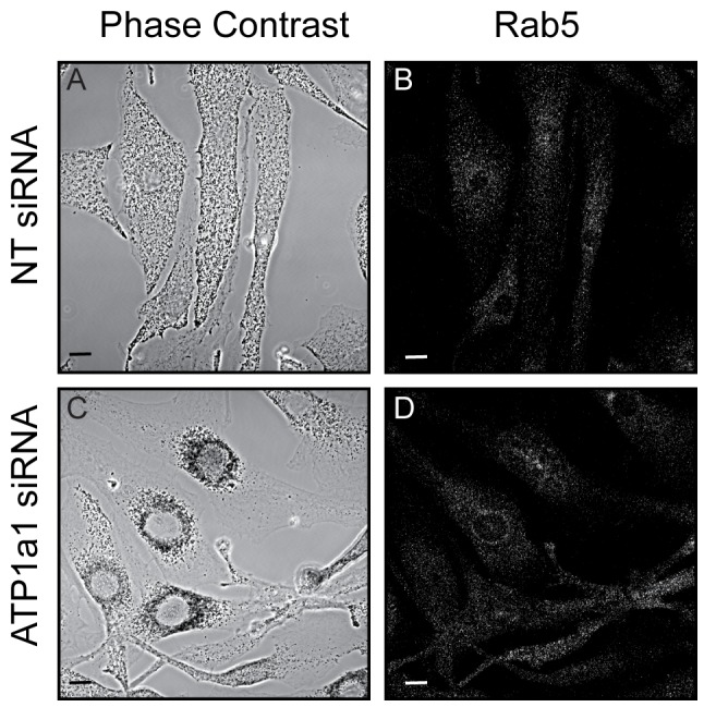
Figure 3. Rab5a-positive vesicle distribution is unaffected following ATP1a1 depletion.

Melan-INK4a cells were treated with either NT or ATP1a1 siRNA and cultured for 72 h. Cells were fixed with PFA, permeabilised and immunolabelled with antibodies to Rab5a. Phase contrast panels show melanosome distribution (A and C), panels B and D show Rab5a staining. Scale bar represents 10 µm.
