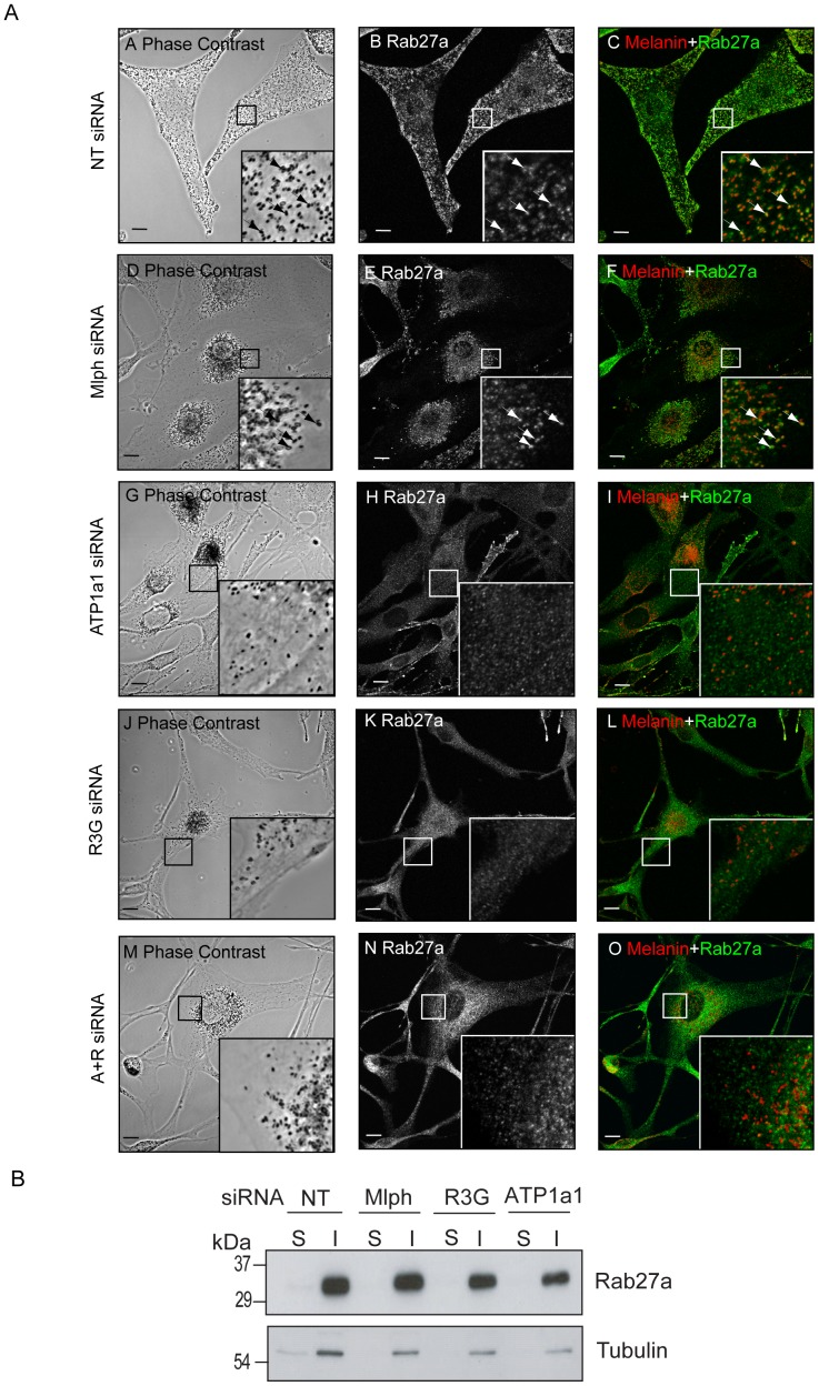
Figure 6. ATP1a1 depletion disrupts endogenous Rab27a targeting to melanosomes.
A) Melan-INK4a cells were plated on coverslips and treated with NT (A–C), Mlph (D–F), ATP1a1 (G–I), R3G (J–L) or ATP1a1+R3G (M–O) siRNA for 72 h before repeating the treatment. After a total of 7 days, cells were fixed with PFA, permeabilised and immunolabelled with an antibody to Rab27a. Phase contrast panels show melanosome distribution (A, D, G, J, M); panels B, E, H, K, N show Rab27a localisation. In the merge panels (C, F, I, L, O) the pigment is inverted and pseudo-coloured red to aid co-localisation with the green Rab27a signal. Insets are a higher magnification of the boxed area. Arrows indicate co-localisation. Scale bar represents 10 µm. B) Melan-INK4a cells were treated with NT, Mlph, R3G or ATP1a1 siRNA and cultured for 3 days. The PNS was then separated into the insoluble (I) and soluble (S) fraction by centrifugation at 100,000×g. Rab27a and Tubulin partitioning were assessed by immunoblotting with the antibodies indicated.

