
Figure 2. IFNγ-induced expression of IRF-1.
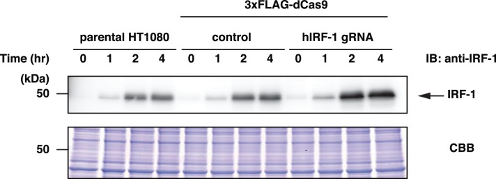
HT1080 and its derived cells were stimulated with 100 ng/ml of IFNγ for indicated time intervals. Nuclear extracts were subjected to SDS-PAGE and immunoblot analysis with anti-IRF-1 Ab. CBB staining is shown as a protein loading control.