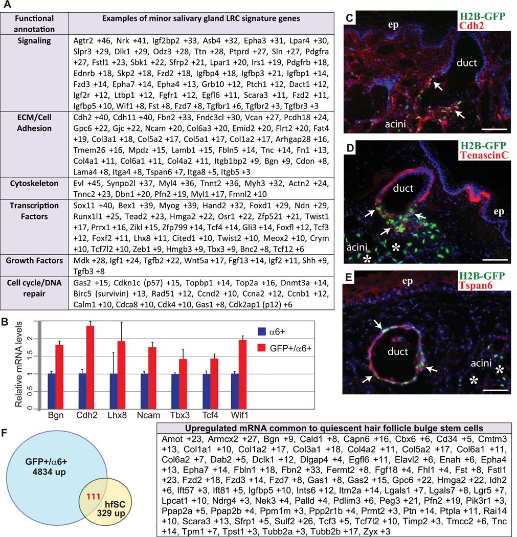
Figure 3. Transcriptional profiling of minor SG LRCs.
Functional annotation of genes up-regulated (>2.5×) in minor SG LRCs (A). Real-time PCR confirmation of selected signature genes of the minor SG LRCs obtained in array analysis (B). Side view sections of soft palate of mice after 4-week chase with H2BGFP (green) and 4’,6’-diamidino-2-phenylindole (DAPI) (blue), and indirect immunofluorescence with antibodies (Abs) for Cdh2, TnC, Tspan6 as indicated (Red) (C–D). Comparisons of minor SG LRCs and hair follicle LRCs profiles. Venn diagram showing similarities between the minor SG LRCs and hair follicle LRCs molecular signature (F). Table in (F) highlights several of the key similarly expressed genes in minor SG LRCs and hair follicle LRCs. Scale bar: C–E 50µm.

