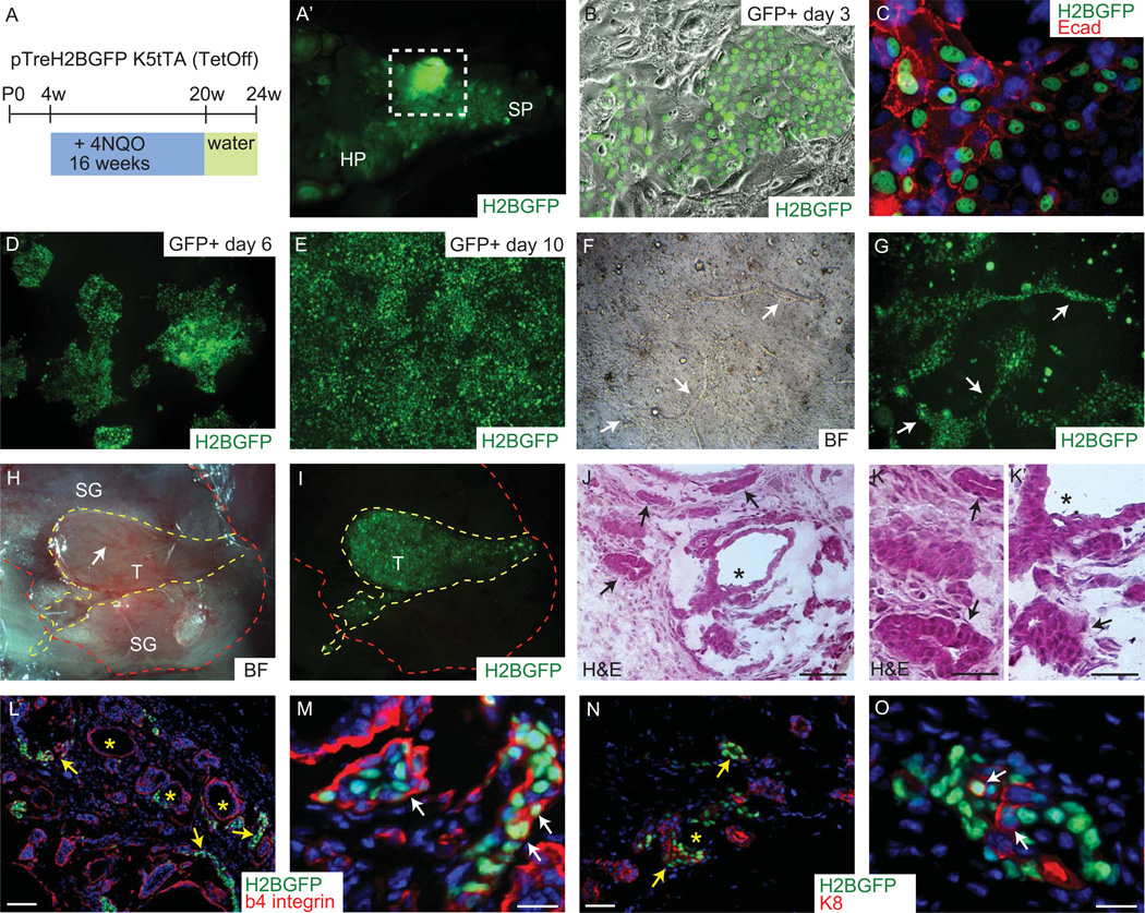
Figure 6. Tumorigenic potential of minor SG basal layer cells.
Schematic of pTreH2BGFP/K5tTA mice with 4NQO treatment (A). View of mouse soft palate with developed tumors in the minor SG (A’). Tumor cells were placed in culture and formed epithelial colonies (B–E), as indicated by E-cadherin presence (C). Minor SG tumor cells formed tubular-like structures when placed in 3D matrigel (arrows in F–G). Injected minor SG tumor cells into the gland of NOD.Cg mice formed secondary tumors that are highly vascularized (arrow in H) and could be traced due to the GFP label (I). Shown is H&E for tumor morphology (J, K and K’) and epifluorescence of H2BGFP (green) and 4’,6’-diamidino-2-phenylindole (DAPI) (blue), and indirect immunofluorescence with antibodies as indicated (Red) (L–O). Scale bar: L and N 100µm; M and O 20µm. Arrows in J, K, L, and N indicate tubular-like structures, arrows in K’ show solid pattern and asterisk in J, K’, L and N indicate microcystic structures. Arrows in (M) indicate basal layer localization of GFP positive cells; arrows in (O) indicate K8 expression in GFP positive cells. Abbreviations: HP, hard palate; SP, soft palate; SG, salivary gland; T, tumor.

