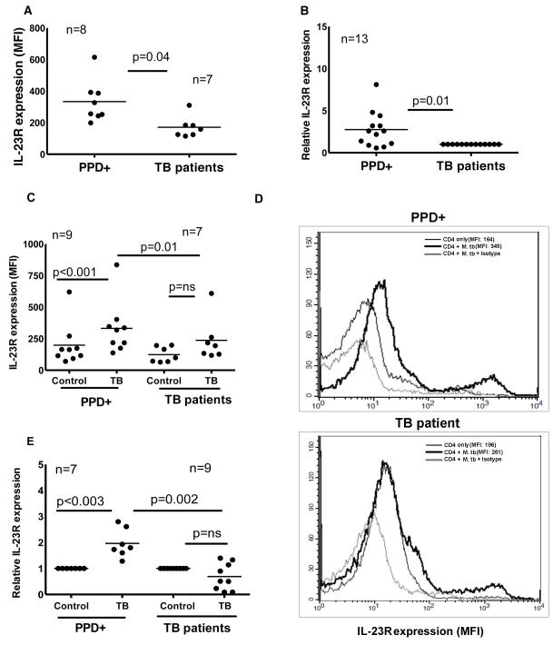
Figure 3. IL-23R expression by T cells from healthy PPD+ donors and tuberculosis patients.
A. IL-23R expression by CD4+ cells. PBMC from 8 PPD+ individuals and 7 tuberculosis patients were isolated from the blood and stained with anti-CD4-FITC and anti-IL-23R-APC. CD4+ cells were gated and the mean fluorescence intensity (MFI) of IL-23R was measured by flow cytometry. Seven independent experiments were performed. B. IL-23R mRNA expression by freshly isolated CD4+ cells. CD4+ cells from freshly isolated PBMC of 13 PPD+ individuals and 13 tuberculosis patients were isolated by immunomagnetic selection. RNA was extracted and reverse transcribed to cDNA, and IL-23R mRNA was quantified by real-time PCR, relative to expression of GAPDH. Each point shows the mean of triplicate determinations. Thirteen independent experiments were performed. C. IL-23R expression by M. tb-stimulated CD4+ cells. CD4+ cells and CD14+ cells from freshly isolated PBMC of 9 PPD+ individuals and 7 tuberculosis patients were isolated by magnetic beads conjugated to anti-CD14 or anti-CD4 obtained from Miltenyi Biotech. CD4+ cells and CD14+ cells were cultured at a 9:1 ratio, with or without γ-irradiated M. tb H37Rv. After 96 h, surface staining for CD4 and IL-23R was performed using anti-CD4-FITC and anti-IL-23R-APC. CD4+ cells were gated and the mean fluorescence intensity (MFI) of IL-23R was measured. Seven independent experiments were performed. D. A representative figure for panel C is shown. IL-23R expression by CD4+ cells of medium alone (thin black line), CD4+ cells of γ-irradiated M. tb H37Rv cultured cells (thick black line) and IL-23R isotype antibody staining on CD4+ cells of γ-irradiated M. tb H37Rv cultured cells (thick light line). E. IL-23R mRNA expression by M. tb-stimulated CD4+ cells. CD4+ cells from PBMC of 7 PPD+ individuals and 9 tuberculosis patients were cultured as in panel C. After 96 h, RNA was extracted and reverse transcribed to cDNA, and IL-23R mRNA was quantified by real-time PCR, relative to expression of GAPDH. In all panels, the thin lines show mean values. Seven independent experiments were performed. Paired and unpaired t tests were performed. Mean values, p values and number of donors (n) in each panel is shown.

