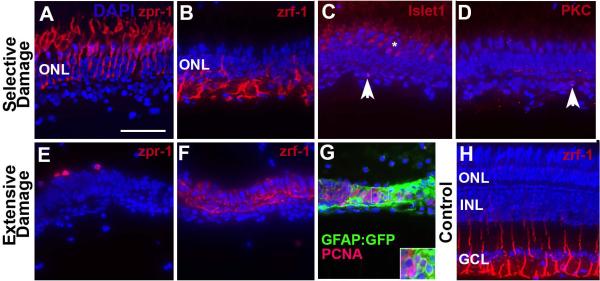
Figure 1.
Characterization of surviving retinal cells following selective or extensive damage. A–D. Retina was subjected to selective damage and processed for sectioning and indirect immunofluorescence three days after the ouabain injection. Cone photoreceptors (A; zpr-1+) appear uninjured; Müller glia (B; zrf-1+) are present but show unusual morphologies; rarely islet1+ cells (C; horizontal cells or debris, arrowhead) are present; and even more rarely PKCα+ cells (D; bipolar cells or debris, arrowhead) survive the injury. E–F. Retina was subjected to extensive damage and processed for sectioning and indirect immunofluorescence four days after the ouabain injection. Cone photoreceptor debris (E; zpr-1+) is present; Müller glia (F; zrf-1+) are present but show highly unusual morphologies. G. GFAP:GFP transgenic retina subjected to extensive damage and processed for confocal imaging of GFP, PCNA, and DAPI. Clusters of PCNA+/DAPI+ nuclei are surrounded by GFAP+ cytoplasm. H. Undamaged adult zebrafish retina labeled with zrf-1 to show normal morphological appearance of basal glial processes. Scale bar (applies to all) = 25 μm.

