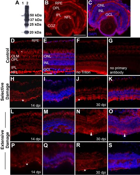
Figure 7.
Distribution of Hh protein in undamaged and regenerating zebrafish retinas. A. Western blotting results using the anti-Shh antibody from AnaSpec (AnaSpec, Fremont, CA; #55574). Lane 1, proteins derived from zebrafish embryos; lane 2, molecular size ladder. Proteins in lane 1 likely correspond to unprocessed (50kDa) and processed (20kDa) Hh protein. B. In a 4 day old larval zebrafish eye, anti-Shh antibody stains the retinal pigmented epithelium (RPE), the outer plexiform layer (OPL), the inner plexiform layer (IPL), the nerve fiber layer (NFL), and extracellular regions of the circumferential germinal zone (CGZ). C. Same as (B), but with DAPI staining to show locations of nuclear layers. OLM, outer limiting membrane; ONL, outer nuclear layer; INL, inner nuclear layer; GCL, ganglion cell layer. Scale bar in B (applies to A–C) = 100 μm. D. Undamaged adult zebrafish retina, showing Hh staining in the retinal pigmented epithelium (RPE), the inner and outer plexiform layers (IPL, OPL), the nerve fiber layer (NFL), and cone photoreceptor inner segments (*). E. Same as (D), but colabeled with DAPI to show positions of outer nuclear layer (ONL), inner nuclear layer (INL), and ganglion cell layer (GCL). F. Undamaged control retina, immunofluorescence performed in the absence of Triton X-100. G. Undamaged control retina, experiment performed in the absence of primary antibody. H. Retina subjected to selective damage of inner retinal layers, sampled at 14 dpi (days post-injury). The most intense staining corresponds to processes (asterisks) associated with an emerging NFL. RPE and photoreceptors are also stained. I. Same as (H) but colabeled with DAPI. J. Retina subjected to selective damage, sampled at 30 dpi. Staining pattern is similar to that of undamaged control retinas, with prominent staining of the NFL, although the retina remains histologically disorganized. K. Same as (J) but colabeled with DAPI. L. Retina subjected to extensive damage of all retinal layers, sampled at 14 dpi. Regenerating plexiform layers are stained, but NFL staining is not evident. M. Same as (L) but colabeled with DAPI. N. Retina subjected to extensive damage, sampled at 30 dpi. Staining pattern includes regions of apparent ectopic Hh (arrowheads), associated with histological abnormalities. O. Same as (N) but colabeled with DAPI. P. Retina subjected to extensive damage, sampled at 14 dpi, view is of a rare region containing Hh staining at the vitreal surface (asterisk). Q. Same as (P) but colabeled with DAPI. R. Retina subjected to extensive damage, sampled at 30 dpi, view is of a rare region containing Hh staining at the vitreal surface (asterisk). S. Same as (R) but colabeled with DAPI. Scale bar in E (applies to D-N) = 25 μm.

