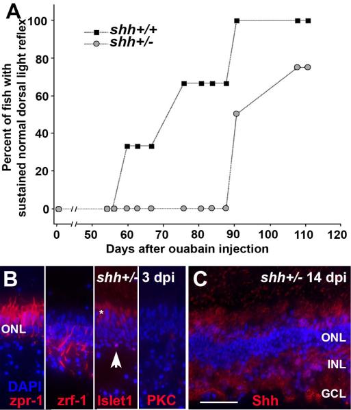
Figure 8.
A. Significantly delayed recovery of normal dorsal light reflex (DLR) following unilateral selective retinal lesion in shhat4+/− zebrafish as compared to their shha+/+ siblings. Difference between groups, p=0.001 (logistic regression with repeated measures). B. Characterization of surviving retinal cells in shhat4+/− retinas sampled at 3 dpi following selective damage. Cone photoreceptors (zpr-1+) are uninjured; Müller glia (zrf-1+) are present but show unusual morphologies; some horizontal cells (islet1+; arrowhead) are present but pyknotic; bipolar cells (PKC+) very rarely survive the injury. Asterisk (*) indicates autofluorescence of photoreceptor inner segments. C. Retinas of shhat4+/− zebrafish subjected to selective damage display evidence of histological recovery, with widespread laminar fusions, at 14 dpi; compare to (Fimbel et al., 2007). Also at 14 dpi, the distribution of Hh protein (in red, with DAPI nuclear stain in blue) in shhat4+/− retinas is diffuse (compare to Fig. 7). Scale bar in B (applies to B–C) = 25 μm.

