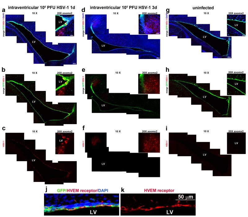
Figure 1. Susceptibility of NPCs present in the ependymal and subventricular cell layers of the lateral ventricle to HSV-1 infection.
Nestin-GFP transgenic mice were inoculated with 105 PFU HSV-1 or PBS as control in the right lateral ventricle of the brain. At 1 (panels a-c) or 3 (panels d-f) days post infection, the mice were perfused with 4% PFA, their brains removed and sectioned (50μm), and subsequently processed. Confocal images of representative brain slices were used to reconstruct the whole margins of the lateral ventricle (LV). GFP-expressing NPCs are depicted in green, HSV-1-expressing cells are shown in red and DAPI-stained nuclei in blue. Brain slices from uninfected mice (panels g-i) served as controls for HSV-1 antigen expression. Note, co-localization of GFP and HSV-1 antigen at day 1 pi (panel b) within the ependyma and SVZ. By day 3 pi, the HSV-1 antigen expression is primarily expressed in the SVZ proximal to the ependyma. The HSV-1 entry receptor, HVEM, was found constitutively expressed within the ependymal and SVZ of naïve uninfected mice, shown in red, co-localizing with GFP+ expressing cells (green) (panels j-k). LV: lateral ventricle. This figure is representative of n=3 mice/time point or uninfected controls.

