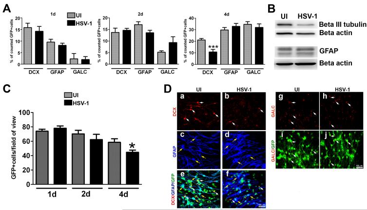
Figure 5. Differentiation patterns of NPCs following infection with HSV-1.
NPCs were seeded on Matrigel matrix covered plates in EGF free media and 3 h later, the cultures were infected with HSV-1 (MOI = 10−4) or left uninfected (UI). At the indicated time pi (day 1-3), cultures were fixed and immunostained for neuron (DCX), astrocyte (GFAP) and oligodendrocyte (GALC) markers (A, C, D). The bar graph in A shows the percentage of positive cells for each marker ±SEM comparing infected to UI cultures (***p<0.005 by paired T test comparison between UI and HSV-1 infected for DCX). B. Representative blots from protein lysates probed with antibodies against β III tubulin (neuronal marker) and GFAP 4 days pi. The graph in C shows the averaged absolute number of cells per field of view ± SEM comparing HSV-1 and UI cultures at each time point. To quantify cell numbers, 5-7 fields of view were counted within a sample for each cell marker in 3 independent experiments (at least 6 total replicates per measurement), *p<0.05 comparing the UI to HSV-1infected group at day 4 pi by paired T test comparison. D. Representative confocal images show a selective decrease in the percentage (and number, data not shown) of DCX-positive cells 4 days pi. White arrows indicate DCX+ or GALC+ cells and yellow arrows indicate GFAP+cells.

