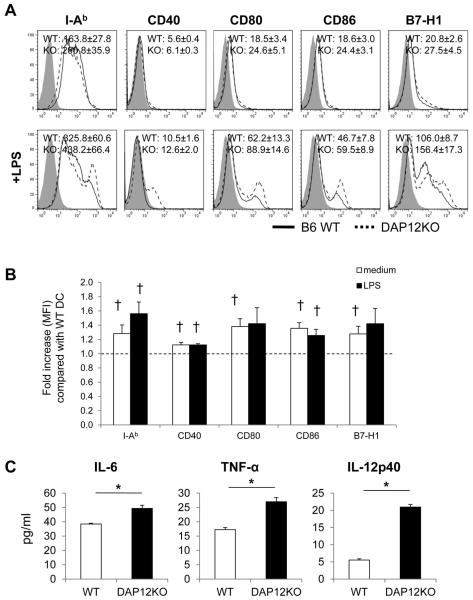
Figure 1. DAP12−/− mouse (C57BL/6; B6) liver myeloid (m) dendritic cells (DC) express elevated levels of cell surface MHC class II (IAb), co-stimulatory and co-regulatory molecules and secrete increased levels of pro-inflammatory cytokines.
(A) flow cytometric analyses of mAb-stained cells wild-type (WT) or DAP12−/− liver mDC cultured overnight in the absence or presence of LPS. Grey profiles indicate isotype controls. Representative data are shown, together with the mean fluorescence intensity (MFI) +/−1SD for each molecule. (B) Fold increase in MFI for each molecule expressed by DAP12−/− compared with WT liver mDC. (C) Concentrations of IL-6, TNFα and IL-12p40 in culture supernatants of unstimulated or LPS-stimulated WT and DAP12−/− liver mDC. Data shown are means +/−1SD obtained from n=4 independent experiments.

