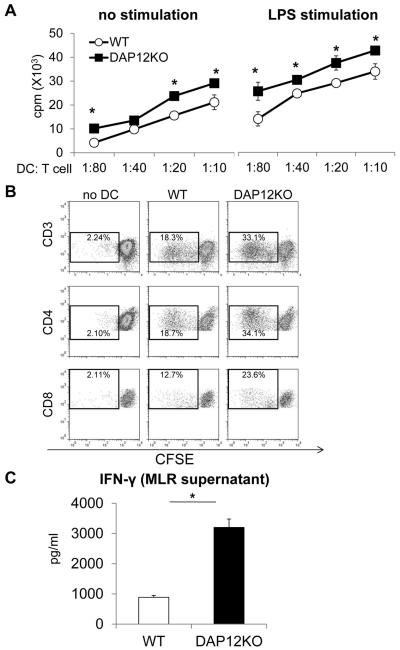
Figure 2. Enhanced in vitro T cell allostimulatory activity of DAP12−/− compared with WT liver mDC.
Liver mDC were cultured with normal bulk allogeneic BALB/c T cells for 72 hr as described in the Materials and Methods. (A) Extent of T cell proliferation induced by unstimulated or LPS-stimulated DC at various DC:T cell ratios determined by thymidine incorporation. *, p< 0.05. (B) Extent of CD4 and CD8 T cell proliferation induced by WT or DAP12−/− liver mDC at a DC:T cell ratio of 1:10 determined by CFSE-MLR. (C) levels of IFNγ detected in MLR supernatants following T cell stimulation by WT or DAP12−/− liver mDC. *, p<0.01. Data are from n=4 independent experiments.

