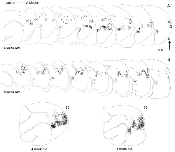
Figure 2. Serial reconstructions of retrogradely labeled cells in the visual cortex of juvenile ferrets.
The semi-tangential sections are arranged serially (lateral=left). Black dots represent retrogradely labeled cells. A, B: Feedback label in every fourth section in a 4 week old (A) and an 8 week old (B) ferret. C, D: Superimposed and aligned images of the serial sections from the reconstructions shown in A and B to reveal the complete pattern of label. At 4 weeks postnatal, the overall label pattern resembles that seen in the adult. The sparse labeling anterior to area 21 and dorsal to the suprasylvian area (Ssy) is located in areas PPr and PPc. A: anterior, D: dorsal.

