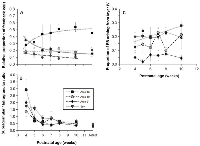
Figure 3. Developmental changes in the areal and laminar distribution of feedback cells in the visual cortex of juvenile ferrets.
A: Proportion of feedback projections to area 17 arising from areas 18, 19, 21, and Ssy as a function of age. Before eye opening, Ssy makes the largest feedback contribution to area 17; by 6 weeks postnatal, area 18 feedback input is largest. B: Ratio of the proportion of feedback to area 17 arising from supragranular to that of infragranular layers in each area. The supragranular contribution to feedback declines synchronously in all extrastriate areas from 4 to 6 weeks. C: The proportion of feedback arising from layer IV in each area changes little in this period, except for feedback from area 18 whose layer IV contribution increases. Adult mean values are plotted on the right for comparison in panels A and B. Error bars represent ±SEM

